Fluorescence analysis technology is a powerful analytical tool widely used in clinical testing, biological research, agricultural science, food and environmental science. It is an important application of multi-function microplate readers, such as TECAN (M1000, M200, etc.). Thmeral (Varioskan Flash), Bio-tek (Synergy 4, etc.), MD (M2, M5) can be applied to fluorescence detection.
1. Overview
At room temperature, most of the molecules are at the lowest vibrational level of the ground state. The molecules in the ground state absorb energy (light energy, chemical energy, electrical energy or thermal energy) and then transition to the excited state. The excited state is unstable and will decay to the ground state very quickly. The form of light emits energy, a phenomenon known as "lighting phenomenon." Molecular luminescence includes fluorescence, phosphorescence, chemiluminescence, bioluminescence, and the like. When it is illuminated by light, the light that disappears immediately when the light is cut off is called fluorescence. When the light is cut off, the light is gradually weakened so that the disappearance is called phosphorescence. The chemical energy of chemical reaction is absorbed and the light is called chemiluminescence, and the energy is converted into light radiation. For bioluminescence.
Since the fluorescent material has different molecular fluorescence and atomic fluorescence, the molecular fluorescence is the band spectrum, and the atomic fluorescence is the line spectrum. The so-called fluorescence is molecular fluorescence. The substance can be qualitatively and quantitatively analyzed by measuring the characteristics and intensity of the emitted fluorescence.
2. Fluorescence detection technology
2.1 Fluorescence intensity (FI)
The fluorescence intensity is proportional to the concentration of the fluorescent substance, which is the basis for quantitative analysis by fluorescence analysis. It is widely used in biology for biomacromolecule quantification, enzyme activity analysis, fluorescence immunoassay, cytological analysis (cell proliferation, cytotoxicity, cell adsorption, etc.) and intermolecular interactions.
2.1.1 Apoptosis detection
The Caspase family plays a very important role in mediating apoptosis, with Caspase-3 being a key executive molecule that functions in many pathways of apoptotic signaling. Caspase-3 is normally present in the cytosol in the form of a zymogen (32KD), which is activated in the early stages of apoptosis. The activated Caspase-3 consists of two large subunits (17KD) and two small subunits ( The composition of 12KD) cleaves the corresponding cytosolic nucleus substrate, which ultimately leads to apoptosis. However, in the late stage of apoptosis and death cells, the activity of caspase-3 was significantly decreased.
A short peptide Z-DEVD-AMC coupled with a fluorescent substance was designed. In covalent coupling, AMC cannot be excited by fluorescence, and short peptides are hydrolyzed to release AMC, and free AMC can be excited to emit fluorescence (Fig. 1). Based on the magnitude of the released AMC fluorescence intensity, the activity of caspase-3 can be determined to reflect the extent to which Caspase-3 is activated.
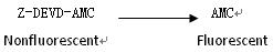
Figure 1. Caspase-3 hydrolysis Z-DEVD- AMC
2.1.2 Detection of cytotoxicity
In vitro cytotoxicity studies have important implications for the detection of new biologically derived or synthetic cytotoxins as well as routine clinically relevant assays. The cell membrane non-permeable nuclear dye Propidiu m iodide can penetrate the damaged cell membrane. The higher the fluorescence density, the more damaged cells are reflected.
2.1.3 calcium flow detection
Fura-2, indo-1, and Quin-2 are Ca 2+ fluorescent indicators that can sensitively reflect changes in intracellular calcium concentration. When combined with calcium ions, the maximum excitation wavelength changes, and the intensity and combination of emission fluorescence The concentration of Ca 2+ has a quantitative relationship.
2.2 Fluorescence Polarization (FP)
In 1926, Perrin first described the theory of fluorescence polarization. Fluorescent molecules in solution can absorb and release the corresponding polarized fluorescence when exposed to polarized light. If the fluorescent substance is at rest during excitation, the emitted light will retain the original excitation light. Polarization, if it is in motion, the plane of polarization of the emitted photoelectric polarization will be different from the polarization of the original excitation light. This is the phenomenon of fluorescence polarization, the interaction of fluorescent molecules with other factors, such as mutual binding or repulsion; The nature of the environment, such as the viscosity of the solution, temperature, etc., may affect the emission plane of the emitted light emitted by this fluorescent factor. Therefore, techniques based on fluorescence polarization can be used to study interactions between molecules in life sciences, such as receptor ligand binding assays, DNA-protein binding assays, SNP assays, and enzyme activity assays.
Fluorescence polarization analysis requires less sample, high sensitivity, sub-nanadene range, good repeatability, easy operation, safer and more reliable, and does not generate harmful radioactive waste during the experiment. In addition, fluorescence polarization is True homogeneous, allowing real-time detection (kinetic detection), insensitive to concentration changes, is the best solution for homogeneous detection (with no washing step in between).
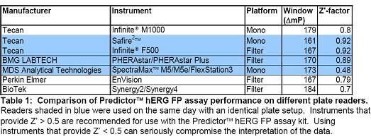
A variety of microplate readers are currently available for fluorescence polarization detection. Invitrogene uses PredictorTM hERG to perform fluorescence polarization analysis on a variety of microplate readers (Table 1).
2.3 Time-Resolved Fluorescence (TRF)
When performing fluorescence measurements, the sensitivity of using conventional chromophores for fluorescence detection is severely degraded due to background fluorescence signal interference. Most background fluorescence signals are present for a short period of time, so the use of long decay lifetime markers minimizes transient fluorescence interference.
Time-resolved fluorescence is the use of rare earth elements as a marker. The structure of the electron cloud of rare earth trivalent ions limits the migration of electrons to a certain extent. The decay period of the fluorescence of such elements is usually very long, thus eliminating background fluorescence. The interference greatly increases the sensitivity of the test (Table 2). Another benefit of using rare earth elements as labels is that the excitation light and the emitted light peak Stoke displacement are large. This eliminates the interference of the excitation and scattered light. At the same time, the excited fluorescent band is extremely narrow, and the emission peak of the fluorescence is very sharp, which allows the instrument to be adjusted in a very narrow wavelength range, greatly reducing the background. Various interferences.
Fluorophore | Fluorescence lifetime (ns) |
Non-specific fluorescent background | 1 to 10 |
Human serum albumin | 4.1 |
globulin | 3.0 |
Cytochrome C | 3.5 |
Fluorescein isothiocyanate (FITC) | 4.5 |
Dansyl chloride | 14 |
Rare earth chelate | 10 3 to 10 6 |
Table 2. Common fluorescence team fluorescence lifetime
Time-resolved fluorescence sensitivity, high specificity, good stability, easy preparation of markers, good detection repeatability, short operation procedure, suitable for ultra-micro analysis of biology and medicine, such as hormone detection, detection of viral hepatitis markers , targeted cell marker detection and drug screening.
2.4 Fluorescence Resonance Energy Transfer (FRET)
Fluorescence resonance energy transfer was first discovered by Perrin in the early 20th century. In 1948, Foster founded the theoretical principle, which refers to the process of transferring energy between a fluorescent energy donor and a receptor through dipole-dipole coupling. The transfer is non-radioactive, and there are three main conditions for FRET: (1) the donor and the acceptor are close enough (1 to 10 nm); (2) the emission spectrum of the donor overlaps with the excitation spectrum of the acceptor; (3) The dipole of the donor and the acceptor has a certain spatial orientation, which is the condition of dipole-dipole coupling.
Fluorescence resonance energy transfer is often used to study the interaction between molecules, such as protein interactions, antigen-antibody binding, receptor-ligand binding, and in the membrane, because of the distance between the donor and the acceptor. Studies on reactions, ion channels, etc. have also been applied. The FRET fluorescent probe-labeled peptide chain was added to a double-layer membrane on a solid surface and detected by fluorescence bleaching recovery (FRAP) imaging technique to provide a new method for studying transmembrane helix dimerization. The cytoplasm was labeled with FRET and its gated effect on the P2X ion channel was detected using time-resolved techniques.
The long-acting fluorescent substance such as Eu is used as a donor to perform fluorescence resonance energy transfer. After the excitation light is extinguished, the receptor can still have a longer energy decay time, higher energy transfer efficiency, and a longer detectable interaction distance. 100-200nm, time delay detection, reduced background noise and improved sensitivity
3. Summary
Fluorescence method is sensitive, accurate, and compatible. It can use fluorescent molecules to specifically mark the target substance, greatly reduce the signal interference of impurities, have many fluorescent characteristic parameters, wide dynamic linear range, can detect living cells, and sensitivity ratio spectrophotometry. It is 2 to 4 orders of magnitude higher, and it has great advantages for sensitive and accurate detection of trace and trace drugs. In summary, fluorescence method, as a highly sensitive analytical method, combined with other technologies, has a broader development prospect.
Multi Enzyme,Feed Additives Enzymes,Feed Enzymes,Nsp Enzyme
Tangshan Finely Animal Care Co.,Ltd , https://www.faczyme.com